Field Identification of Foliar Cotton Diseases Can Oftentimes be Based on Canopy Location
Related Articles
- Rice Variety Trial Results For 2010, Plus Rice Research Report 0
- Evaluation of Peanut Prescription Rx Program in Mississippi 0
- Fertilizing Cotton with Poultry Litter 5
Latest Tweets
Over the last several years it has become increasingly important to consider specific foliar diseases in cotton. The best way to consider the specific disease present is to break the cotton canopy into thirds (bottom third, middle third, upper third). Even though breaking the canopy into thirds is a general way to consider the diseases outlined below there are always exceptions to these rules. Remember, the environment tends to dictate the presence of specific diseases and can also specifically dictate how much each of the diseases outlined below influences the cotton crop.

Symptoms associated with the potassium-associated leaf spot complex as they appear in the upper canopy.
Foliar potassium deficiency-associated leaf spots
Present almost exclusively in the UPPER most canopy. In addition, and more generally, lesions are not typically observed on plants except in the upper most nodes and do not extend into the middle or lower canopy. Leaves on plants do not always present a nutrient deficiency. Even though the specific pathogens are associated with a nutrient deficiency, the deficiency is not in the soil, it is present in the plant. Lesions tend to be brown to gray in the middle with a maroon margin. The specific organisms involved are almost exclusively opportunistic and need the deficiency within the plant for the lesion to occur. In severe cases, the foliage can develop a nutrient deficient response and defoliation can result. Multiple organisms can cause these lesions and are most notably a specific species of Cercospora or Stemphyllium. Telling the two organisms apart is not so important as is the proper diagnoses regarding the underlying cause.
Prior experience suggests that all cotton varieties are likely susceptible to this particular set or organisms.
Management of this particular disease situation requires managing the potassium deficiency in the plant and not attempting to manage the disease with a fungicide. However, with many of the early-maturing cotton varieties, heavy fruit load can influence the source to sink relationship and once nutrients are shifted towards boll production the leaves can lose nutrients and the leaf spot issues are generally observed.

Symptoms associated with bacterial blight. Clockwise from upper left: water-soaked lesions as observed on the bottom of the leaf, systemic vein infection as observed in non-conducive environments, black to ashy-colored lesion on petiole, and lesions on bracts.
Bacterial blight
Bacterial blight can oftentimes be observed throughout the canopy. However, and more frequently, bacterial blight will first be observed in the middle of the canopy. Normally the bacterium that causes bacterial blight does not like the environment in the upper canopy and in those instances the symptoms associated with bacterial blight can appear quite different.
Bacterial blight can produce lesions on all plant parts that each appear different. The lesions generally associated with bacterial blight on leaves appear as small, angular lesions and are best observed by considering the appearance on the top and bottom of the leaf surface. Lesions on the upper leaf surface appear dark and those on the underside of the leaf generally appear dark with severe water-soaking around the lesion margins. However, lesions on leaves are best observed early in the day when dew is present and the water-soaking is most severe. In situations when the environment is not favorable for the disease the lesions can coalesce along the vein. Lesions on the petiole and stem appear darkened and generally circle the petiole or stem. The lesions that develop on the bract appear similar to what might occur if you held an old style fountain ink pen to the plant tissue and allowed the ink to run. In situations where the lesions are observed on bolls they will initially appear round and water-soaked. As the lesions mature on bolls they may turn dark in color and in most cases are invaded by secondary saprophytes which will generally rot the majority of the boll.
Numerous varieties are susceptible to bacterial blight. Information regarding the specific reaction of varieties can be confirmed by checking with the specific seed company or accessing the bacterial blight response information at: https://www.cottoninc.com/wp-content/uploads/2018/01/Management-of-Bacterial-Blight-of-Cotton.pdf
Management of bacterial blight once the disease is observed is all related to the subsequent season. Choosing a resistant variety in field situations where the disease has been observed is important. In addition, management in the subsequent season can be influenced by tillage. Burying residue can limit the presence of the bacterium surviving the winter on residue that remains on the soil surface (especially in mild climates). Honestly, the best way to manage bacterial blight is to rotate out of cotton for a year in situations where the disease has been observed. But, I realize that is not always an economically feasible situation for all cotton farmers.

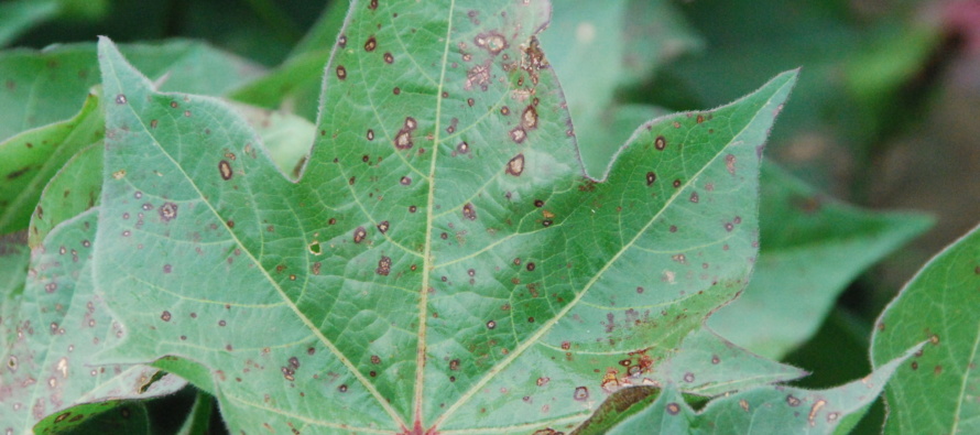
Target spot of cotton. Note defined lesions with concentric rings without the presence of a yellow halo (which is not always present).
Target spot
Target spot is most generally observed in the lower canopy. However, environment dictates target spot location within the canopy. In years when excessive rainfall occurs that keeps the middle canopy wet target spot can result in a high level of disease in the mid-canopy as well as defoliation. The symptoms associated with target spot in the lower canopy consist of gray to brown lesions with concentric rings. Some lesions may contain a yellow halo around the lesion itself; however, not ALL lesions contain the yellow halo and this is not one of the better diagnostic features associated with target spot. Symptoms associated with target spot are generally detected after the canopy has closed as the organism requires the increase in humidity as well as reduced light for widespread infection to occur.
Even though target spot tends to be observed in the lower canopy, the disease can result in different lesion types expressed in the mid-to-upper canopy. However, when the symptoms differ in the upper canopy the disease has been present in the lower canopy for some time so do not focus on lesions in the upper canopy and assume that target spot is occurring their first. In the upper canopy, if the environment is conducive enough for the disease to move into that part of the canopy, lesions may not develop concentric rings and will tend to be smaller in size (e.g., 1/8 inch as compared to ½ inch in size).
Based on observations made during the 2017 season (see: https://www.mississippi-crops.com/2018/01/27/cotton-target-spot-2017-lucedale-ovt-disease-rating-results/) the bulk of the cotton varieties planted in the MS cotton production system appear susceptible to target spot.
Management of target spot appears to be a complex issue. A large number of university-conducted target spot fungicide trials from the Mid-south suggest that automatically timed fungicide applications (generally at first flower) are not economically beneficial. Trials conducted in MS for the past 3-4 seasons suggest that fungicides are not economically beneficial for managing the disease. However, with that said, the disease has generally occurred at or a little beyond 5NAWF. Moreover, even in situations where a fungicide is applied defoliation will STILL be observed. While there is some truth to statements regarding a reduction in defoliation as a result of some specific fungicide products defoliation will still be observed in the low-to-mid canopy.

Bacterial blight can develop a yellow halo that oftentimes will be confused with target spot.
Lesion mimics
Keep in mind that several diseases can be confused based on the appearance of the lesions and where those occur within the canopy. As with any specific lesion or lesion-type, an herbicide can produce a lesion that appears similar to any of the diseases outlined above. One specific example that comes to mind would be the lesion that occurs as a result of Liberty injury on non-Liberty cotton. Quite regularly, the lesion that develops as a result of Liberty-injury has a stark yellow halo around the lesion itself.
Specific lesion mimics include:
-herbicide injury that can appear like multiple diseases (potassium-associated lesions)
-bacterial blight with a yellow halo that appears similar to target spot (see image)
In addition, in some cases the lesions associated with bacterial blight can develop a yellow halo that oftentimes can be confused with target spot. Scouting for the presence of blight can aid in the proper diagnosis since as mentioned above, not all target spot lesions will develop a yellow halo.


Let me tell You a sad story ! There are no comments yet, but You can be first one to comment this article.
Write a comment